
A structured breast screening programme that is fast and highly accurate, combining specialist medical expertise with the latest digital technology.
In Italy, more than 50,000 women are diagnosed with breast cancer every year. The encouraging news is that mortality continues to fall. In 2025, against roughly 56,000 new diagnoses, five-year survival reached 86%, rising to 90% when the disease is caught in its early stages. Part of the credit goes to scientific advances in treatment, but above all to the spread of prevention, to mammography and to screening programmes extended to the whole population, which make it possible to identify the disease early and to intervene more effectively. It is also thanks to prevention that almost a million women in Italy have today come through one of the hardest trials of their lives.
ASC, a medical centre dedicated to cancer prevention and early detection, has chosen to make a real contribution to this challenge, so crucial to women’s health. It does so by bringing together, in line with its long-held approach, the most advanced digital diagnostic technology and leading medical specialists.
Our breast health pathway offers a structured screening programme that is fast and highly accurate, designed to detect any breast conditions at an early stage.
At ASC, specialist expertise and the latest technology work together to deliver care that is thorough, tailored to you and reassuring at every stage of the diagnostic process.
The pathway includes:
State-of-the-art 3D imaging that reconstructs the breast in thin layers for highly precise early detection.
A first-line diagnostic examination, particularly suited to assessing glandular tissue and dense breasts.
Reporting and direct clinical assessment with the breast radiologist, to complete and interpret the diagnostic picture.
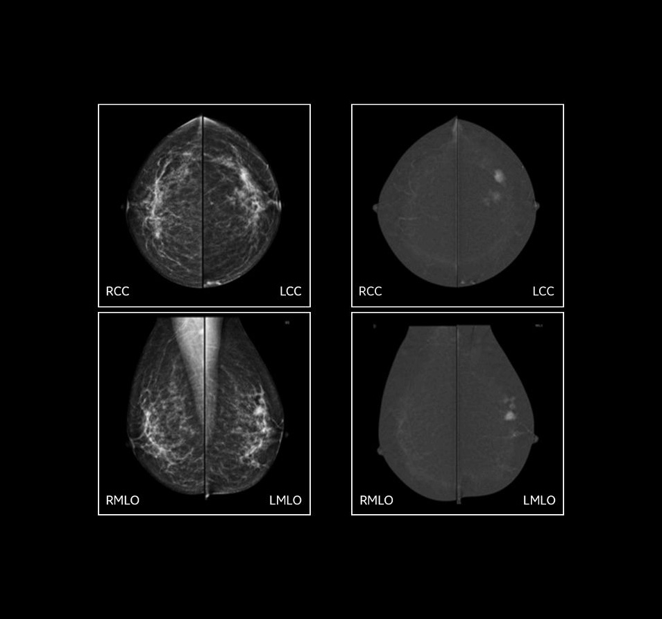
Mammography with tomosynthesis, also known as three-dimensional (3D) mammography, is an evolution of conventional mammography in the early detection of breast cancer. The main difference lies in the quality of the images acquired. Where conventional (2D) mammography produces a limited number of flat, two-dimensional images, tomosynthesis captures many projections from different angles, reconstructing the breast in very thin layers. This reduces the effect of overlapping tissue and significantly improves diagnostic precision, that is, the ability to identify even very small lesions.
During a tomosynthesis examination, the X-ray tube moves in an arc while taking a series of sequential exposures. The acquisition is then processed by dedicated software, which reconstructs the breast in sections around one millimetre thick, giving an extremely detailed representation of the area being examined.
The images reconstructed in this way allow an accurate assessment of the fine structures of the breast, free from unwanted tissue overlap and unaffected by any high tissue density: small lesions and suspicious densities can therefore be identified with greater accuracy and precision.
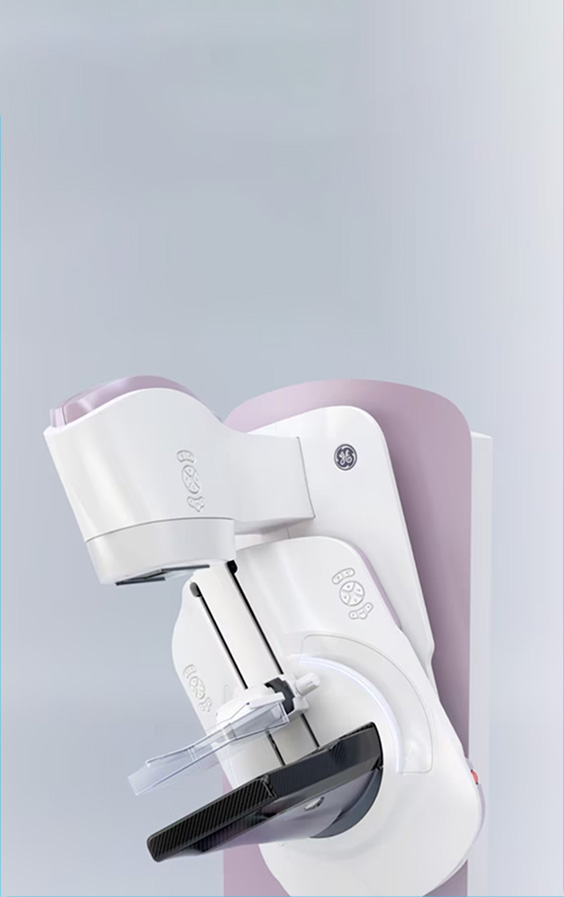
The examination is very similar to a conventional mammogram and involves gently compressing the breast, which is needed to reduce its thickness and obtain high-quality images.
The main difference lies in the movement of the X-ray tube, which rotates in an arc, capturing a large number of low-dose images from different angles. Dedicated software then processes these projections to generate three-dimensional images in very high definition.
Thanks to the three-dimensional reconstruction of the breast, the ability to identify invasive lesions increases by up to 41%, revealing even small lumps or changes that might remain hidden in two-dimensional images.
(that is, fewer “false positives”)
Better visualisation of the tissue significantly reduces the need to recall patients for further tests, because it makes it easier to tell normal anatomical overlap from genuinely suspicious areas.
The breast compression required is often lighter and better tolerated than with conventional mammography.
Today’s technology keeps X-ray exposure extremely low, entirely comparable to that of a standard digital mammogram.
Breast ultrasound is a first-line examination that complements mammography and is particularly suited to assessing glandular tissue and denser breasts.
It is a non-invasive diagnostic examination that uses ultrasound (high-frequency sound waves that are harmless to the human body and its tissues) to examine the breast tissue and the lymph nodes in the armpit on both sides.
It can accurately distinguish the nature of lumps and cysts, completing the diagnostic picture and providing an even more precise assessment.
The pathway is completed by the clinical breast examination: a thorough and completely painless assessment carried out by a doctor, without the use of any equipment.
During the examination the doctor gathers information and examines the breasts by looking at them and palpating them carefully, in order to identify or rule out any changes, conditions or risk factors.
Our breast health pathway begins with mammography with tomosynthesis, followed by breast ultrasound together with the clinical breast examination, and ends with the report and a consultation on the results.
Thanks to our dedicated breast pathway, reports are prepared quickly and are available as soon as the examinations and consultation are complete, simply and securely. Qualified staff The team of radiography professionals who carry out mammography at ASC is made up of highly qualified specialists, with long-standing experience and particular care for the person in front of them. The breast early-detection pathway is entrusted to a radiologist with specific expertise in breast imaging, who oversees every stage of the diagnostic process directly. As well as reporting on the mammogram, the specialist personally performs the breast ultrasound and the clinical examination, ensuring a fully integrated assessment.
Every woman is different, with her own characteristics and needs. That is why each pathway is shaped around the person, through direct discussion between the woman herself, the radiographer and the specialist, adapting the examinations to her particular clinical needs. Our priority is to provide accurate, reliable diagnoses, so that every woman can go through the diagnostic process with complete peace of mind and a clear understanding of what is happening.
The spaces at our centre are designed to offer calm, privacy and comfort, helping to ease the anxiety that diagnostic tests can often bring. Every room is intended to welcome the person with discretion and care, in a setting built around wellbeing, listening and respect for confidentiality.
If you notice a lump, a change in the skin or shape of your breast, or any discharge from the nipple, book an appointment without waiting for your annual check-up. A prompt assessment means the situation can be clarified quickly and, if necessary, acted on straight away.
If you have no symptoms, a breast examination is recommended as a routine yearly check. As a general guide, it is combined with a breast ultrasound before the age of 40 and with a mammogram from that age onwards. If there is a family history of breast cancer, your doctor may advise starting these checks earlier.
Complete breast screening package
Payment can be made by debit card, credit card or bank transfer.
A special rate
for those who also book the Diffusion Whole Body scan
Payment can be made by debit card, credit card or bank transfer.
Our doctors are experienced specialists with proven expertise in breast imaging.
A transparent early-detection pathway, with clear answers at every stage.
At ASC you are a guest, not a patient: you are at the centre of everything we do, welcomed in comfortable surroundings.
The Senographe Pristina is a full-field digital mammography system, designed to provide a complete solution for breast imaging.
We're here for you
We'll guide you through your booking every step of the way. We'll get back to you as soon as possible with all the information you need to go ahead.
Booking form


